New Nuffield Health research shows more than 40 per cent of women who’ve had a mammogram say they found it painful and nearly 15 per cent avoid having a breast screening due to fear of discomfort.
Shockingly, more than one in 10 women (12 per cent) say they have avoided having a breast check, despite finding a lump.
SoGlos speaks to Fiona Court, breast surgeon at Nuffield Health Cheltenham Hospital and Jo Evans, senior mammographer at Nuffield Health Cheltenham Hospital, to find out what a breast screening is really like.
What actually happens when someone comes in for a breast screening?
Coming in for a breast screening is really important. In the UK a woman is diagnosed with breast cancer every 10 minutes.
While that’s a big number, when we catch cancers early, the chances of survival and a good outcome are much higher. So it’s really important to regularly self-check, take up screenings when offered and above all make sure you check out any lumps, bumps or abnormalities.
Thankfully, while the screening process used to be much more invasive, the technology has moved on a long way, so the process should never put you off coming.
Typically, after you’ve been checked in at the front desk, you’ll be ushered through to the imaging department, where you’ll be met by a mammographer.
In most organisations (including in Nuffield Health), this will be a female and she’ll ask you simple questions about any previous screening, family history and any current concerns you have.
If anyone is putting off their screening appointment, I would let them know all ladies are hesitant to remove their clothing and feel vulnerable. However, all our mammographers are women who understand breast anatomy and the majority have had a mammogram themselves, so can help to allay any fears.
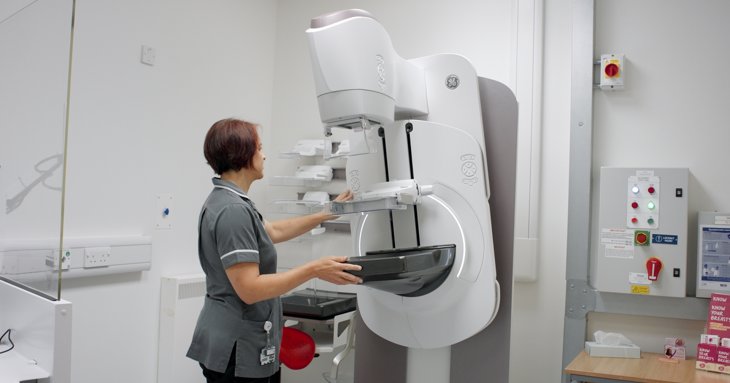
She’ll then explain exactly what the mammogram involves – undressing to the waist, having four X-ray images taken, two from the top and two from the side. You’ll stand in front of the machine and lean in and they will help you get into the right position.
Previously this could be quite uncomfortable but the machines are much better at moving around women and the sharp edges have been rounded out in most machines.
A plastic compression paddle is then applied slowly by the mammographer to even out the breast tissue and to keep the breast still while the images are taken.
The mammogram images are reviewed by the mammographer and normally two highly trained breast radiologists also. Typically, the results are shared within two weeks (if it’s a health screening) or if there are concerns around symptoms, this may even be shared immediately.
Whatever the outcome, the team will always explain the next steps. If anything is found that needs further investigation, the consultant and specialist nurse will explain that clearly for you.
How long does the procedure typically take and how many images are taken?
The technology is now much faster and provides clearer images, so the mammogram will take approximately 20 minutes including the explanation and imaging.
Typically, four images is enough for the consultant to pinpoint areas within the breast, using two dimensions to work out the location of any changes.
One of the biggest concerns is pain – how would you describe the sensation of a mammogram to someone who’s never had one?
The compression feels similar to a blood pressure cuff but is controlled at all times by the mammographer to ensure patient comfort – most women describe the compression as a pinch.
We have several sizes of compression paddle to accommodate all sizes of breast tissue and each mammogram only lasts a few seconds with compression.
The good news is with the newer machines, the amount of compression needed is much less, therefore less uncomfortable.
Why does compression need to be used and how much pressure is actually applied?
Compression makes the breast tissue thin enough for the x-rays to pass through and give an image. If the breast is too thick, then the x-rays will not pass through and the image will be all white.
Lumps such as cancers or cysts are more dense than the surrounding breast tissue and this is what we’re looking for.
If the compression is not intense enough then early signs may be missed.
So what about younger women and women with dense breasts?
In younger women – those under 40 – mammograms are generally not performed as the breast tissue is much denser and lumps are less likely to show up. This is why we do not generally offer mammogram to younger women.
However, with the use of our 3D system using Tomosynthesis technology, we can image the breast using multiple x-ray images at different angles. These images are then combined to create a 3D picture of the breast to allow easier reporting and to improve the accuracy of detecting changes.
The technology is improving all the time and we check for anything concerning, but the most important message is, if you find something, get it checked.
Don’t worry about how it’s done, leave that to us. Just make sure you come in and let us do the rest.
Are there any common myths or misconceptions about mammograms you’d like to clear up?
Some women have heard mammograms cause cancer due to the radiation exposure but this really isn’t true.
The radiation dose is very small, similar to the background radiation we are exposed to in our normal lives – equivalent to a few transatlantic flights.
The new unit here at Cheltenham is the state of the art GE Pristina and offers both 2D and 3D mammograms. With this machine the radiation is even lower, as we can take a 3D image using the same dose as a 2D one.
Another myth is women with implants cannot have a mammogram, that somehow the compression will rupture the implant. You absolutely can have a one. They are sometimes more difficult to read and implants may hide some parts of the breast but we have our tricks and techniques to get around this, so it is always worth having a mammogram.
You may be asked to sign a disclosure in the case of a rupture but this is highly unlikely. If there is a rupture, it’s nearly always because there was a defect and the implant was already damaged.
Some women think breast ultrasound is an alternative to mammogram however this also isn’t the case. Breast ultrasound is used to look at a specific part of the breast, looking for specific changes and symptoms.
They don’t act as a screening, i.e. checking the whole breast, so they're not able to detect many early signs of breast cancer.
What would you say to someone who is putting off their screening because they’re afraid it will hurt or be embarrassing?
If anyone is putting off their screening appointment I would let them know all ladies are hesitant in one way or another.
Whether that’s concern over the process, whether it will hurt or concern about removing their clothing and feeling vulnerable.
All the mammographers at Nuffield Health Cheltenham are women and most have been through it themselves. They are here to support you and can help to ease any fears.


.jpg?width=690&height=361&rmode=pad&bgcolor=ffffff&quality=85)


.jpg?width=432&height=227&rmode=pad&bgcolor=ffffff&quality=85)


